
 |
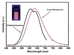
| Luminescence of photodynamic particles with X-ray activation--Courtesy of the Journal of Biomedical Nanotechnology |
Researchers at the University of Texas at Arlington found that nanoparticles they were studying for radiation detection in the security arena could produce a toxic byproduct able to damage cancer cells.
In what is called a photodynamic therapy method, which delivers light to a tumor as a cancer treatment, UT physicist Wei Chen found that copper-cysteamine, or Cu-Cy, nanoparticles lost energy over time when combined with X-ray exposure, which led to slower growth of nearby cancer cells. Chen published the research in the Journal of Biomedical Nanotechnology, to be released in August.
Often photodynamic therapy is induced with a near-infrared laser, but Chen found that X-ray exposure allowed for deeper tissue penetration. And the Cu-Cy nanoparticles can work on their own without other photosensitizers, like other luminescent particles used previously for this purpose.
The team tested the method on human breast and prostate cancer cells and found in one case that a tumor stayed "virtually the same size" over 13 days while an untreated tumor grew to about three times its original size.
"This new idea is simpler and better than previous photodynamic therapy methods," Chen said in a statement. "You don't need many steps. This material alone can do the job. It is the most promising thing we have found in these cancer studies and we've been looking at this for a long time."
Co-author Lun Ma added: "Photodynamic therapy is limited to treat superficial diseases such as skin cancer because current photosensitizers cannot be activated using high energy irradiations that are able to deeply penetrate into tissues. … It may be a breakthrough to bring photodynamic therapy to deep cancer treatment."
- here's the UT-Arlington report
- and here's the abstract